
How to distinguish a creatinine rise during effective decongestion from one that requires a change in treatment.
Clinical bottom line
- Continue diuretics when congestion persists and perfusion is maintained. A mild creatinine rise alone is not a reason to routinely interrupt decongestion. (professional.heart.org )
- An inadequate response may require more effective diuresis, not less. Persistent fluid overload should prompt reassessment of treatment response and potentially escalation. (ESCARDIO )
- Reduce or temporarily withhold treatment when there is convincing hypovolaemia or treatment-related haemodynamic deterioration. A congested patient with hypoperfusion needs urgent specialist assessment—not automatic fluid administration or further diuretic escalation. (Enlighten Publications )
- Interpret creatinine alongside urine output, congestion, perfusion and its subsequent trajectory. Biochemical worsening does not, by itself, establish structural kidney injury. (Scholars@Duke )
This review addresses adults receiving inpatient treatment for acute or decompensated heart failure. Cardiogenic shock and urgent indications for kidney replacement therapy require a separate emergency pathway.
Does a rising creatinine mean the diuretic is damaging the kidneys?
Not necessarily: creatinine reflects filtration, not the mechanism of kidney dysfunction.
During decongestion, haemodynamic changes can reduce glomerular filtration without substantial tubular injury. In the ROSE-AHF biomarker analysis, worsening renal function during aggressive diuresis was not accompanied by increases in measured tubular-injury biomarkers. This supports a distinction between a functional filtration change and structural injury; it does not establish that every creatinine rise during diuresis is benign. (Scholars@Duke )
Congestion itself can also impair kidney function. Therefore, reducing diuresis solely because creatinine has increased may leave an important contributor to renal dysfunction untreated. Conversely, excessive fluid removal can produce intravascular depletion and worsen perfusion. (Enlighten Publications )
The practical question is not simply “Has creatinine increased?” It is “What has happened to the patient while creatinine increased?”
How much of a rise should trigger concern?
Use acute kidney injury criteria to trigger reassessment—not as an automatic diuretic stopping rule.
KDIGO defines acute kidney injury through any of the following:
- Creatinine increase ≥26.5 µmol/L (0.3 mg/dL) within 48 hours.
- Creatinine increase to ≥1.5 times baseline within seven days.
- Urine output <0.5 mL/kg/hour for six hours. (KDIGO )
There is no universally validated creatinine increase that establishes either “safe to continue” or “must stop” for loop diuretics. In particular, the often-quoted 30% creatinine threshold associated with renin–angiotensin system treatment should not be repurposed as a loop-diuretic stopping threshold. (Enlighten Publications )
A progressive rise accompanied by falling urine output, loss of diuretic response or evidence of hypoperfusion is more concerning than an isolated increase during otherwise effective treatment. (NCBI )
What should be checked before changing the prescription?
Reassess congestion, perfusion and competing causes of kidney dysfunction together.
Establish whether orthopnoea, jugular venous pressure, oedema and other signs of congestion are improving. Review measured urine output, fluid balance and weight together rather than allowing one measurement to determine treatment. Assess blood-pressure trends, peripheral temperature, mental state and symptoms of hypotension. Lactate is appropriate when hypoperfusion is suspected—not simply because creatinine has risen. (ESCARDIO )
Look for another explanation: sepsis, recent nephrotoxic exposure, urinary obstruction or intrinsic kidney disease. Urinalysis and urine sediment are useful when the pattern is unexplained or atypical; urinary tract imaging is indicated when obstruction is suspected. A recent reliable baseline and the timing of medication changes help establish the trajectory. (KDIGO )
When should diuretics be continued, reduced or stopped?
The decision should follow the clinical phenotype, not the creatinine result in isolation.
| Clinical situation | Diuretic decision | Immediate priority |
|---|---|---|
| Persistent congestion, maintained perfusion and useful diuretic response | Continue, with reassessment | Complete decongestion without causing depletion |
| Persistent congestion and inadequate response, without hypoperfusion | Optimise or intensify treatment | Check response and consider combination therapy |
| Convincing hypovolaemia or over-diuresis | Reduce or temporarily withhold | Correct the cause and reassess haemodynamics |
| Congestion with hypoperfusion, progressive oliguria or unexplained deterioration | Do not automatically escalate or stop all fluid removal | Urgent cardiac and renal assessment |
This is a practical synthesis of heart-failure guidance and inpatient diuretic recommendations, rather than a validated decision score. (professional.heart.org )
An asymptomatic blood-pressure reduction is not equivalent to shock. Equally, oedema does not exclude impaired effective circulating volume. Discordant findings should prompt better haemodynamic assessment rather than reflex prescribing. (professional.heart.org )
What should happen when congestion persists but diuresis is inadequate?
Measure the response early and adjust treatment while reassessing safety.
The 2026 ESC decongestion algorithm uses a spot urine sodium of ≥70 mmol/L at two hours, or urine output of ≥100 mL/hour over the first six hours, as indicators of an adequate initial response. An inadequate response can prompt an increased intravenous loop dose or an additional diuretic, provided the clinical assessment supports further fluid removal. These are response benchmarks—not proof of renal safety. (ESCARDIO )
Urine sodium is useful where available, but it should complement clinical assessment. In PUSH-AHF, natriuresis-guided treatment increased sodium removal without demonstrating a reduction in death or heart-failure rehospitalisation. (Groningen Research Portal )
The DOSE trial also illustrates why creatinine should not be considered alone: higher-dose treatment produced greater fluid removal but more short-term worsening renal function. It did not demonstrate worse subsequent clinical outcomes, although it was not powered to establish long-term safety conclusively. (PubMed Central (PMC) )
Can a second diuretic be added when creatinine is already rising?
Yes, in selected patients with persistent congestion—but combination therapy increases the need for monitoring.
The 2026 ESC guideline supports considering short-term intravenous acetazolamide or oral hydrochlorothiazide in fluid-overloaded patients previously receiving loop diuretics. This is a recommendation to improve decongestion, not evidence that combination treatment protects kidney function. (ESCARDIO )
The ADVOR renal analysis found that acetazolamide increased treatment-period worsening renal function. However, creatinine did not differ between treatment groups at three months, and the observed renal deterioration was not associated with worse clinical outcomes in that analysis. These findings require caution because post-randomisation associations cannot prove that an individual patient’s deterioration is harmless. (Groningen Research Portal )
In CLOROTIC, hydrochlorothiazide improved weight loss but substantially increased the frequency of renal-function deterioration. A subsequent analysis found no statistically significant association between early kidney-function declines and adverse outcomes, but its confidence intervals were wide. (OUP Academic )
Therefore, an additional diuretic is an option for unresolved congestion—not a response to creatinine itself. Check agent-specific renal restrictions and monitor electrolytes and acid–base status; do not extrapolate trial findings to uncontrolled shock or severe, rapidly progressive kidney injury. The broader trial evidence shows a trade-off between more effective decongestion and biochemical adverse effects. (PubMed Central (PMC) )
What monitoring and escalation are required?
Monitor both the effectiveness of fluid removal and evidence of treatment intolerance.
Check renal function and electrolytes at least daily during intravenous diuresis. Twice-daily testing is reasonable in higher-risk patients—for example, those with substantial renal dysfunction, large fluid losses or combination treatment—although this intensified schedule is based largely on expert practice rather than randomised comparisons. Follow urine output, weight, fluid balance and haemodynamics alongside laboratory results. (ESCARDIO )
Progressive oliguria, haemodynamic deterioration or an unexplained renal trajectory should trigger urgent reassessment rather than another routine dose increase.
Ultrafiltration is not the default response to a rising creatinine. In CARRESS-HF, stepped pharmacological treatment preserved renal function better than ultrafiltration, achieved similar weight loss and caused fewer serious adverse events. This does not exclude ultrafiltration for appropriately selected patients with refractory congestion. (Scholars@Duke )
Urgent kidney replacement therapy is determined by life-threatening fluid, electrolyte or acid–base complications and the overall clinical context—not an isolated creatinine threshold. (KDIGO )
When can intravenous treatment be stepped down?
Step down when congestion has adequately resolved and a sustainable oral regimen has been established—not solely when creatinine returns to its previous value.
Avoid both premature withdrawal with residual congestion and continued aggressive fluid removal after the patient has become depleted. Discharge planning should include an oral diuretic strategy, instructions for adjustment and a clearly assigned plan for clinical and laboratory reassessment. Progressive unexplained kidney dysfunction still requires resolution of the diagnostic and management plan. (professional.heart.org )
Bedside decision algorithm
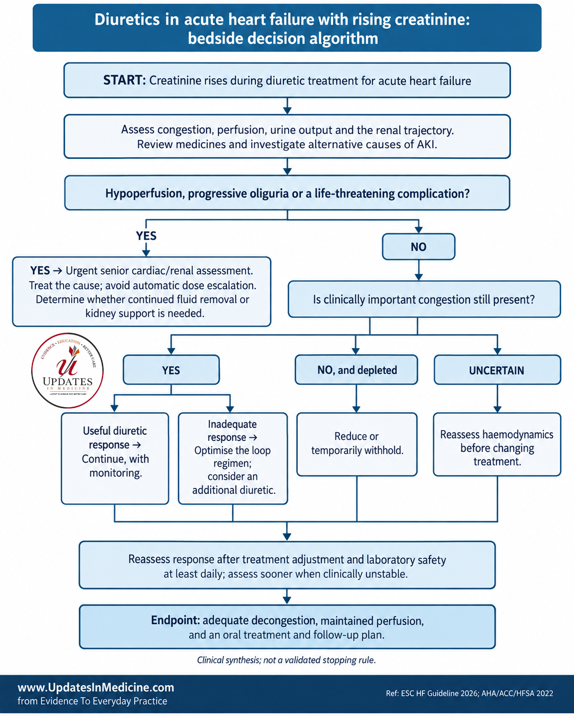
START: Creatinine rises during diuretic treatment for acute heart failure
↓
Assess congestion, perfusion, urine output and the renal trajectory.
Review medicines and investigate alternative causes of AKI.
↓
Hypoperfusion, progressive oliguria or a life-threatening complication?
├─ YES → Urgent senior cardiac/renal assessment.
│         Treat the cause; avoid automatic dose escalation.
│         Determine whether continued fluid removal or kidney support is needed.
└─ NO
↓
Is clinically important congestion still present?
├─ YES
│   ├─ Useful diuretic response → Continue, with monitoring.
│   └─ Inadequate response → Optimise the loop regimen;
│                            consider an additional diuretic.
├─ NO, and depleted → Reduce or temporarily withhold.
└─ UNCERTAIN → Reassess haemodynamics before changing treatment.
↓
Reassess response after treatment adjustment and laboratory safety
at least daily; assess sooner when clinically unstable.
↓
Endpoint: adequate decongestion, maintained perfusion,
and an oral treatment and follow-up plan.
Clinical synthesis; not a validated stopping rule. (ESCARDIO )
Common practice pitfalls
| Pitfall | Better approach |
|---|---|
| “The creatinine rose, so furosemide must stop.” | Determine whether congestion or depletion predominates. |
| “The patient is losing fluid, so the kidney injury must be harmless.” | Reassess perfusion, urine output and alternative causes. |
| “Continuous infusion will prevent renal deterioration.” | DOSE did not demonstrate a renal advantage over intermittent boluses. |
| “More decongestion means a proven survival benefit from the added drug.” | Distinguish improved fluid removal from demonstrated patient-important benefit. |
These distinctions reflect the mechanistic studies, DOSE and contemporary trial synthesis. (Scholars@Duke )
What remains uncertain?
No trial identified in this review directly randomised patients after a creatinine rise to continuing, reducing or stopping diuretics. Much of the decision therefore rests on guideline recommendations, indirect randomised evidence and clinical interpretation.
Neither a universal “acceptable creatinine rise” nor a biomarker-based stopping strategy has been established. Reassuring secondary analyses should not be extrapolated into unrestricted treatment of patients with severe renal deterioration or shock. (Groningen Research Portal )
What to do on the next ward round
Document congestion, perfusion and response before changing the diuretic prescription. State why treatment is being continued, intensified or withheld, and specify what finding will trigger the next change.
The aim is adequate decongestion with maintained perfusion—not an unchanged creatinine at any cost.
Supporting evidence and comparison
Search scope
This focused narrative review used web-mediated searches of PubMed/PMC, official ESC, AHA and KDIGO sources, journal publications and author-hosted research records, with a cutoff of 20 September 2026. It is not a formal systematic review.
The current ESC recommendations were verified against the official 2026 guideline slide set. KDIGO’s 2026 AKI/AKD document remained a draft under preparation, so final AKI recommendations below are attributed to the 2012 guideline. (ESCARDIO )
Guideline comparison
| Source | Relevant recommendation | Strength | Important qualification |
|---|---|---|---|
| AHA/ACC/HFSA 2022 | Do not routinely discontinue diuresis for a mild renal decline or asymptomatic blood-pressure reduction during admission. | Class I, B-NR | Does not justify ignoring hypoperfusion or progressive kidney dysfunction. (professional.heart.org ) |
| ESC 2026 | Intravenous loops for fluid overload; consider acetazolamide or hydrochlorothiazide in previously loop-treated patients. Urine-sodium-guided treatment may be considered. | I A; IIa B1; IIb B1, respectively | Benefits specified are symptom relief and improved decongestion/natriuresis. (ESCARDIO ) |
| Renal Association/British Society for Heart Failure 2019 | Persistent congestion may require continued or increased diuresis; temporarily withhold when hypovolaemic. | Ungraded national guidance | Distinguishes loop-diuretic decisions from renal thresholds used for other HF medicines. (Enlighten Publications ) |
| KDIGO 2012 | Do not use diuretics to treat AKI itself, except for managing volume overload. | 2C | AKI and an indication for fluid removal can coexist. (KDIGO ) |
Interpretation: these recommendations are compatible. Kidney guidance does not prohibit treating congestion in AKI; heart-failure guidance does not endorse diuresis regardless of perfusion.
Comparison of the major studies
Percentage-point differences below are simple differences between the reported event rates.
| Study and population | Comparison | Main finding | Meaning for this decision |
|---|---|---|---|
| DOSE, 2011; 308 patients | Higher- versus lower-dose intravenous furosemide; bolus versus infusion | Worsening renal function: 23% versus 14%, a 9-point increase with higher dosing; greater fluid removal. | A short-term creatinine rise can accompany useful decongestion. Not a trial of continuing versus stopping after AKI. (PubMed Central (PMC) ) |
| CARRESS-HF, 2012; 188 patients with congestion and worsening renal function | Ultrafiltration versus stepped pharmacological treatment | At 96 hours, creatinine changed by +20.3 versus −3.5 µmol/L; weight loss was similar. Serious adverse events: 72% versus 57%. | Supports optimised pharmacological treatment rather than reflex ultrafiltration. (Scholars@Duke ) |
| ROSE-AHF biomarker analysis, 2018; 283 patients | Kidney-function changes during aggressive diuresis versus tubular-injury biomarkers | Worsening renal function occurred in 21.2%, without an accompanying increase in measured tubular-injury markers. | Supports a functional mechanism in some patients; does not exclude injury in others. (Scholars@Duke ) |
| ADVOR renal analysis, 2023; 519 patients | Acetazolamide versus placebo, added to loop treatment | Worsening renal function: 40.5% versus 18.9%, a 21.6-point increase. No between-group creatinine difference at three months. | More effective decongestion can coexist with early renal deterioration. Outcome associations do not prove individual safety. (Groningen Research Portal ) |
| CLOROTIC, 2022/2023; 230 patients | Hydrochlorothiazide versus placebo, added to loops | Weight loss at 72 hours: 2.3 versus 1.5 kg. Impaired renal function: 46.5% versus 17.2%, a 29.3-point increase. | Additional decongestion comes with increased biochemical renal risk; no demonstrated dyspnoea benefit. (OUP Academic ) |
| CLOROTIC renal analysis, 2026; 225 patients with day-two data | Association of early kidney-function decline with outcomes | In the thiazide arm, mortality HR per 30% day-two eGFR decline was 0.96; 95% CI 0.47–1.97. | The estimate was inconclusive; the wide interval does not establish absence of harm. (OUP Academic ) |
| PUSH-AHF, 2023; 310 patients | Natriuresis-guided treatment versus usual care | Sodium excretion at 24 hours: 409 versus 345 mmol. Death/HF rehospitalisation at 180 days: 31% versus 31%; HR 0.92, 95% CI 0.62–1.38. | Supports response-guided fluid removal, but not a proven clinical-outcome benefit. (Groningen Research Portal ) |
| Network meta-analysis, online 2025/print 2026; 25 trials, 7,149 patients | Alternative loop strategies and additional diuretics | Thiazide combinations increased worsening renal function: OR 1.78, 95% CI 1.43–2.21; hypokalaemia: OR 1.69, 95% CI 1.32–2.16. | Combination treatment improves decongestion but requires safety surveillance. Definitions and regimens varied. (PubMed Central (PMC) ) |
Overall synthesis: the evidence argues against reflex withdrawal for every creatinine rise, but equally against labelling all deterioration during diuresis as acceptable. The most defensible approach combines treatment response with perfusion and investigation of competing kidney pathology.
Key claim-to-source map
| Clinical claim | Source location | Evidence basis |
|---|---|---|
| Mild renal decline alone should not routinely interrupt diuresis | AHA/ACC/HFSA 2022, hospitalised-patient treatment recommendations | Class I, B-NR |
| Early urine sodium and urine output can guide escalation | ESC 2026, Figure 15 | Guideline algorithm |
| AKI criteria prompt assessment, not automatic loop withdrawal | KDIGO 2.1.1 and 3.4.2 | Diagnostic criteria and graded guidance |
| More frequent laboratory checks in high-risk diuresis | Michigan Medicine, Q7 | Institutional expert guidance |
| Ultrafiltration is not the default response to cardiorenal deterioration | CARRESS-HF, primary results | Randomised comparison |
The underlying recommendations and their qualifications are detailed above. (professional.heart.org )
References
- European Society of Cardiology. 2026 ESC Guidelines for the management of heart failure. European Heart Journal. 2026. doi: 10.1093/eurheartj/ehag100. Official guideline slide set consulted. (ESCARDIO )
- Heidenreich PA, et al. 2022 AHA/ACC/HFSA Guideline for the Management of Heart Failure. Circulation. 2022;145:e895–e1032. doi: 10.1161/CIR.0000000000001063. (PubMed )
- KDIGO Acute Kidney Injury Work Group. KDIGO Clinical Practice Guideline for Acute Kidney Injury. Kidney International Supplements. 2012;2:1–138. (KDIGO )
- Clark AL, et al. Change in renal function associated with drug treatment in heart failure: national guidance. Heart. 2019;105:904–910. doi: 10.1136/heartjnl-2018-314158. (Enlighten Publications )
- Konerman MC, et al. Michigan Medicine Inpatient Diuretic Guideline for Patients with Acute Decompensated Heart Failure. Michigan Medicine. 2022. (NCBI )
- Cannatà A, et al. Diuretic strategies in acute heart failure: a systematic review and network meta-analysis of randomized clinical trials. European Heart Journal—Cardiovascular Pharmacotherapy. 2026;12:6–14. First online 2025. doi: 10.1093/ehjcvp/pvaf067. (PubMed Central (PMC) )
- Felker GM, et al. Diuretic Strategies in Patients with Acute Decompensated Heart Failure. New England Journal of Medicine. 2011;364:797–805. doi: 10.1056/NEJMoa1005419. (PubMed Central (PMC) )
- Bart BA, et al. Ultrafiltration in Decompensated Heart Failure with Cardiorenal Syndrome. New England Journal of Medicine. 2012;367:2296–2304. doi: 10.1056/NEJMoa1210357. (Scholars@Duke )
- Trullàs JC, et al. Combining loop with thiazide diuretics for decompensated heart failure: the CLOROTIC trial. European Heart Journal. 2023;44:411–421. doi: 10.1093/eurheartj/ehac689. (OUP Academic )
- Ter Maaten JM, et al. Natriuresis-guided diuretic therapy in acute heart failure: a pragmatic randomized trial. Nature Medicine. 2023;29:2625–2632. doi: 10.1038/s41591-023-02532-z. (Groningen Research Portal )
- Ahmad T, et al. Worsening Renal Function in Patients With Acute Heart Failure Undergoing Aggressive Diuresis Is Not Associated With Tubular Injury. Circulation. 2018;137:2016–2028. doi: 10.1161/CIRCULATIONAHA.117.030112. (Scholars@Duke )
- Meekers E, et al. Renal function and decongestion with acetazolamide in acute decompensated heart failure: the ADVOR trial. European Heart Journal. 2023;44:3672–3682. doi: 10.1093/eurheartj/ehad557. (Groningen Research Portal )
- McCallum W, et al. Acute declines in kidney function with thiazide plus loop diuretics versus loop diuretics alone in acute decompensated heart failure. Nephrology Dialysis Transplantation. 2026;gfag070. doi: 10.1093/ndt/gfag070. (OUP Academic )
Evidence limitations: several study results were verified from publisher or author-hosted abstracts rather than complete articles. The full 2026 ESC journal document was not accessible; its official recommendation slides and algorithm were reviewed. The supplied attachment provided the editorial framework rather than clinical evidence.