
Introduction
Infective endocarditis (IE) remains a complex syndrome in which microbiology, cardiac anatomy, embolic risk, haemodynamic status and the feasibility of source control need to be assessed together. The 2026 American Heart Association (AHA) document updates its 2015 scientific statement and covers native-valve and prosthetic-valve endocarditis (PVE), transcatheter valves, cardiac implantable electronic devices (CIEDs), right-sided disease, antimicrobial treatment, surgery and neurological complications.
Importantly, this is an AHA scientific statement rather than a formal clinical practice guideline. The writing group explicitly notes that contemporary AHA scientific statements do not provide formal recommendation classes in the way earlier documents did. Management suggestions therefore combine available evidence with expert consensus in an area where randomised controlled trial data remain limited. Decisions should be individualised, ideally through a multidisciplinary Endocarditis Team.
Why does the Endocarditis Team matter?
The statement places the multidisciplinary Endocarditis Team at the centre of care. Its typical membership includes infectious diseases, cardiology and cardiovascular imaging, cardiac surgery, clinical microbiology, neurology, radiology, dentistry, pharmacy and nursing; addiction medicine is particularly important for people who inject drugs.
The writing group describes this model as optimal for individualised care and notes that multidisciplinary programmes have been associated with lower short-term mortality, shorter time to surgery and greater use of surgery where appropriate. The team also provides a framework for interpreting uncertain imaging, choosing antimicrobial transitions and balancing operative versus neurological risk.
How should suspected IE be diagnosed?
The statement supports the 2023 Duke–ISCVID criteria for contemporary diagnosis. Compared with the 2000 modified Duke criteria, these incorporate molecular pathogen identification, broaden the microbiological definitions relevant to prosthetic material, add multimodality imaging and recognise operative inspection as a major surgical criterion. Validation studies cited by the statement found improved sensitivity, with comparable or modestly reduced specificity depending on the study.
The updated Duke–ISCVID framework also simplifies blood-culture requirements by removing the previous requirement for specific timing and separate venepuncture sites. CCT and ^18F-FDG PET/CT can contribute major imaging criteria.
Which imaging test should come first?
Echocardiography remains first-line. TTE is widely available and non-invasive, with specificity above 90%, but reported sensitivity is approximately 40–66%. TEE has sensitivity and specificity above 90% and is particularly important for prosthetic valves, CIEDs, abscesses and other perivalvular complications.
A high-quality negative TTE may be sufficient to exclude IE in a patient with a native valve and genuinely low pre-test probability. In contrast, TEE is performed in suspected or confirmed PVE and in patients with CIEDs even when TTE is negative or inconclusive. For native-valve disease, TEE is indicated when the post-test probability remains intermediate or high. If suspicion remains high after initially normal or inconclusive echocardiography, TTE or TEE may be repeated within 1 week.
Table: Choosing imaging in IE
| Clinical situation | Practical approach | Important qualification |
|---|---|---|
| Initial suspected IE | TTE first | A negative TTE is less reassuring when pre-test probability is high |
| Prosthetic valve or CIED | TEE | Perform even if TTE is negative or inconclusive |
| Native valve, negative TTE | Escalate to TEE if probability remains intermediate/high | High-quality TTE may be sufficient only with low pre-test probability |
| Suspected PVE with negative/inconclusive echo | Consider ^18F-FDG PET/CT | Particularly useful with prosthetic material |
| Suspected abscess, pseudoaneurysm or periprosthetic extension | CCT is complementary | TEE remains more sensitive for many vegetations |
| Neurological symptoms | Brain MRI preferred | More sensitive than CT for small ischaemic lesions |
| Asymptomatic patient before cardiac surgery | Brain MRI is reasonable | Silent ischaemic emboli alone should not delay surgery |
Figure 1. Imaging pathway when IE is suspected
This editorial pathway summarises the statement's echocardiographic and multimodality-imaging sections. It is not an official AHA algorithm.
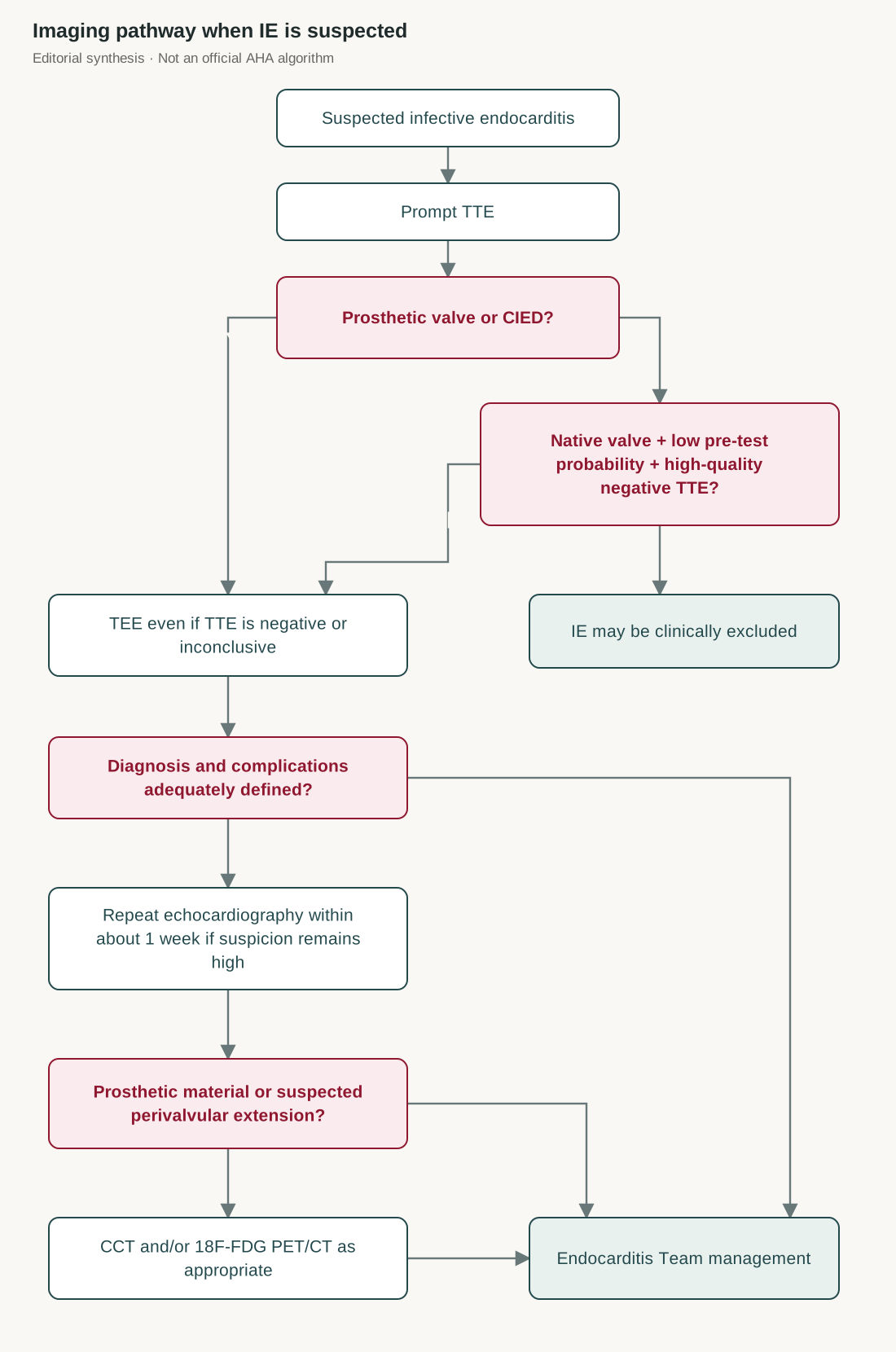
Plain-language equivalent: Start with TTE. Use TEE when prosthetic material or a CIED is present, or when a native-valve patient still has intermediate or high probability after TTE. If the diagnosis remains uncertain despite high suspicion, repeat echocardiography within about a week. In difficult prosthetic or perivalvular cases, add CCT and/or ^18F-FDG PET/CT according to the clinical question.
Source locator: Diagnosis of IE; “The Role of Echocardiography in the Initial Diagnosis of IE”, printed pages e4–e5; “Additional Imaging Modalities in IE”, printed pages e7–e9.
Caption: Editorial synthesis of AHA scientific statement recommendations; not adapted from a single official AHA flowchart.
When should molecular diagnostics be used?
Molecular testing is intended to complement, not replace, conventional microbiology.
If blood and valve cultures are concordantly positive for a typical IE organism, routine molecular testing can generally be deferred. When feasible, the statement suggests storing a pre-antibiotic plasma specimen for possible metagenomic sequencing if blood cultures remain negative at 72 hours.
If an atypical organism is isolated from blood but valve cultures are negative, molecular testing of collected valve tissue is reasonable. In blood culture-negative IE, molecular testing can be applied to valve tissue alongside standard culture and histopathology.
Where does advanced imaging add most?
^18F-FDG PET/CT is particularly useful in PVE when echocardiography is negative or inconclusive but clinical suspicion remains high. Studies cited in the statement report PVE sensitivities above 80–90%, with similar specificity. Its sensitivity in native-valve endocarditis (NVE) is much lower, so routine use for NVE diagnosis is generally not advised. Whole-body PET/CT may also identify metastatic infection.
CCT is particularly valuable for periprosthetic or perivalvular complications such as abscesses and pseudoaneurysms and can assist operative planning. White blood cell SPECT/CT can be considered when suspicion remains high, echocardiography is negative or inconclusive and FDG-PET/CT is unavailable.
Transcatheter-valve disease deserves special caution: device-related artefact reduces the sensitivity of both TTE and TEE in TAVR-associated IE, so multimodality imaging may be required.
How should antimicrobial treatment be structured?
Intravenous antimicrobial therapy remains the cornerstone of the initial phase. The continuation phase may consist of ongoing intravenous treatment, partial oral therapy (POT) in selected patients, or long-acting injectable lipoglycopeptides in selected circumstances.
The statement does not suggest that every patient who improves clinically should switch to oral treatment. Selection criteria are deliberately narrow.
Table: When can partial oral therapy be considered?
| Checkpoint | Source criterion |
|---|---|
| Causative organism | Streptococci, Enterococcus faecalis, methicillin-susceptible Staphylococcus aureus or coagulase-negative staphylococci |
| Initial IV treatment | ≥10 days of appropriate IV antimicrobial therapy |
| If valve surgery occurred | ≥7 days after surgery before transition |
| Clinical response | Clinically stable; no fever for ≥3 days |
| C-reactive protein | <25% of peak value or <20 mg/L |
| Leucocyte count | <15 × 10⁹/L |
| Echocardiography | TEE within 2 days before transition |
| TEE requirements | No progression, abscess formation or new indication for surgery; document vegetation size and characteristics |
Table 5 also excludes POT in circumstances including body mass index >40, suspected reduced gastrointestinal absorption, a concomitant infection requiring intravenous treatment and reduced adherence. The POET population included relatively few PVE cases and very few people who inject drugs, limiting generalisability.
The majority of writing-group members favoured the two-active-agent strategy studied in POET, but consensus was not reached on dual versus monotherapy. POT is described as an option for appropriately selected patients rather than an intrinsically inferior or “second-line” treatment.
What about long-acting agents?
Dalbavancin and oritavancin may provide an alternative continuation strategy when daily intravenous treatment is difficult or prohibitive, or when POT is unsuitable. This should not be interpreted as equivalent evidence to established regimens: the statement highlights limited evidence, concern about relapse—particularly in non-operated prosthetic-valve E. faecalis IE—and emerging glycopeptide/lipoglycopeptide resistance.
Which antimicrobial practices need particular attention?
| Clinical issue | AHA scientific statement position | Qualification |
|---|---|---|
| Gentamicin in staphylococcal PVE | Writing-group consensus: risk outweighs potential benefit | Evidence is largely observational rather than definitive RCT evidence |
| Rifampin in staphylococcal PVE | No consensus | Discuss with the Endocarditis Team |
| Penicillin-susceptible E. faecalis IE | Combination treatment supported | Ampicillin/amoxicillin plus ceftriaxone is favoured by the writing group |
| Staphylococcal NVE followed by valve repair/replacement during treatment | Continuing monotherapy appears reasonable | Based on retrospective, non-randomised evidence; consensus reached |
| S. gallolyticus or E. faecalis IE | Colonoscopy is reasonable to evaluate for colonic malignancy | Consensus statement rather than graded recommendation |
When should surgery be considered?
Surgery complements, rather than replaces, antimicrobial therapy. Broad indications include valve damage causing heart failure, invasive infection with abscess, perforation or fistula, difficult-to-eradicate organisms, failure of antimicrobial therapy, PVE and large vegetations threatening immediate or recurrent embolisation.
Once the indication for surgery during active IE has been established, early surgery within 48–72 hours is favoured, unless neurological complications require a different timing strategy. Surgery aims to remove embolic and infectious sources, drain abscesses, remove infected or necrotic material and restore cardiac integrity and valve function.
For right-sided IE, indications are less firmly defined. The statement lists persistent bloodstream infection, continued septic pulmonary embolisation, pulmonary abscess after 7 days of antibiotics or a right-sided vegetation >2 cm. Valve repair is preferred to replacement where feasible. Percutaneous mechanical aspiration may be considered for selected high-surgical-risk patients in experienced centres.
Figure 2. Timing of surgery after a neurological complication
This algorithm summarises the statement's approach where a patient with IE already has a surgical indication and then has a neurological event.
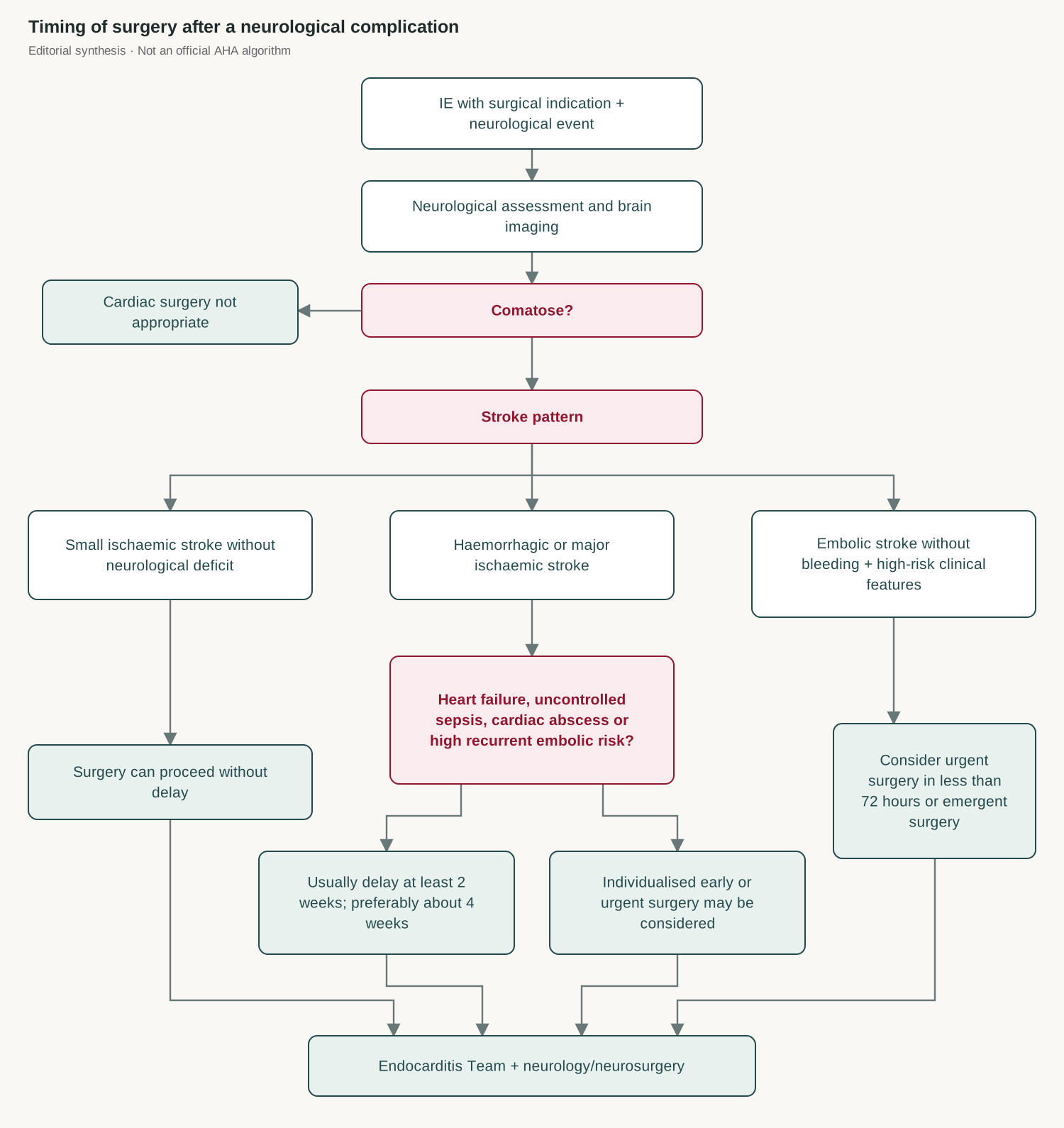
Plain-language equivalent: A small ischaemic stroke without neurological deficit does not necessarily delay indicated surgery. Major ischaemic or haemorrhagic stroke usually leads to a delay of at least 2 weeks and preferably about 4 weeks. However, acute heart failure, uncontrolled sepsis, cardiac abscess or a high risk of recurrent embolism may justify earlier intervention. For embolic stroke without intracranial bleeding and these high-risk features, urgent surgery within 72 hours or emergent surgery should be considered. Large or mobile vegetations >10 mm, mitral involvement—particularly the anterior leaflet—and staphylococcal IE increase recurrent embolic concern. CT/CT angiography or MRI/magnetic resonance angiography should be used to exclude a mycotic aneurysm before cardiothoracic surgery in this setting.
Source locator: “Optimal Timing of Cardiovascular Surgery After Stroke”, printed page e19.
Caption: Editorial synthesis of the AHA scientific statement's neurological and surgical timing recommendations; not an official reproduced algorithm.
How long should antibiotics continue after valve surgery?
One of the most practically important sections concerns how to count treatment when surgery occurs before completion of antimicrobial therapy.
Table: Antimicrobial duration after valve surgery
| Situation | Suggested approach | Important qualification |
|---|---|---|
| Surgery before antimicrobial course is complete | Complete the intended 4–6-week total course, counting pre-operative treatment days | Give at least 14 days post-operatively |
| Completed treatment before surgery, no evidence of active infection and valve cultures negative | No additional post-operative antibiotics needed | Consensus reached |
| Valve culture or molecular test positive after surgery | Positivity alone does not determine duration | Molecular detection of DNA does not necessarily indicate viable organisms |
| Metastatic infectious focus or other complexity | Individualise and consider longer treatment | Endocarditis Team decision |
The practical message is that surgery does not automatically restart a fresh 4–6-week antibiotic course. The planned total is continued, with pre-operative days counted, provided at least 14 days are given after surgery.
What follow-up imaging is needed?
Echocardiography should be repeated whenever clinical status changes during admission or after discharge. TTE is usually performed after completion of antimicrobial therapy to establish a new anatomical and functional baseline; residual thickening or vegetation-like abnormalities need clinical interpretation and do not alone establish persistent infection.
After valve surgery, TTE is routinely performed at 6–12 weeks. Longer-term imaging depends on residual valve disease, prosthesis type and clinical course; in complex surgery, TEE at around 3 months may be considered.
Clinical takeaway
The 2026 AHA scientific statement moves IE care towards an integrated model: diagnose using contemporary Duke–ISCVID criteria, escalate beyond TTE when pre-test probability or prosthetic material warrants it, use molecular and advanced imaging selectively, reserve oral transition for carefully stabilised patients, and treat surgery as timely source control rather than something that must wait until antibiotics are completed. Throughout, the statement repeatedly emphasises individualised Endocarditis Team decision-making, reflecting both the complexity of IE and the limitations of the available evidence.
Full reference
DeSimone DC, Marks L, Dayer MJ, Esquer Garrigos Z, Messika-Zeitoun D, O’Gara PT, Pettersson GB, Bucholz EM, Andrews LS, Fowler VG Jr, Miro JM, Baddour LM; on behalf of the American Heart Association Council on Lifelong Congenital Heart Disease and Heart Health in the Young; Council on Clinical Cardiology; Council on Cardiovascular and Stroke Nursing; and Council on Cardiovascular Radiology and Intervention. Infective endocarditis: diagnosis, antibiotic therapy, and management: a scientific statement from the American Heart Association. Circulation. 2026;154:e•••–e•••. doi: 10.1161/CIR.0000000000001466.